Getting connected with our brain
The brain is responsible for our experience of, and acts as the interface between, the self and the outside world. Everything we think, feel, remember and dream is written by a precisely-interconnected community of ~100 billion brain cells. Have you ever wondered where the different types of neurons in your brain originate from? Or how these brain cells then find their way to connect with other cells, up to a metre away in our body? These answers can be found in the developing brain, which arises from the microscopic, but miraculous, embryo.
Creation of our brain
Very early in human development, the embryo consists simply of three fundamental cell layers: outer ectoderm (becomes outer-body parts e.g. skin/hair/teeth); middle mesoderm (develops into muscles, bones and blood vessels); and inner endoderm (forms our inner-body compartments e.g. gut/lungs). That’s most of our body covered, but where does our brain come from?
A little later, part of the ectoderm is instructed to change its fate. In the embryo, ‘stem’ cells decide what final cell type (e.g. skin/muscle/neuron) they will become by responding to signals from their surrounding environment. In a process of known as ‘Neural Induction’, ectoderm stem cells, located at the back of the embryo, are instructed to become brain stem cells by a signal aptly-named ‘Noggin’. These brain stem cells then roll up to form a ‘Neural Tube’, which will develop into the brain and spinal cord.
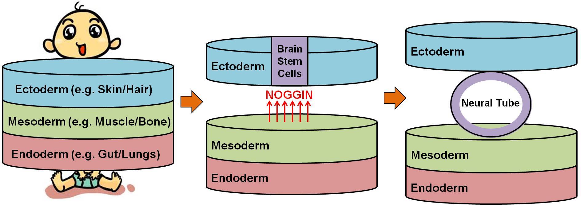
Embryonic Development of Neural Tube from Ectoderm.
Generation of diverse neurons
The relatively uniform ‘Neural Tube’ generates thousands of different types of brain cells by responding to specific combinations of signals. The exact composition of these instructive signals is determined by the location, and time, at which these brain stem cells decide to turn into neurons. Signals come from many sources, typically neighbouring cells, but there are a number of signal ‘centres’ in the ‘Neural Tube’. To gain an appreciation of how this works, we will focus on the spinal cord where there are competing signal centres at its ‘roof’ and ‘floor’.
Signals from the ‘roof centre’ instruct neighbouring brain stem cells to become ‘sensory’ neurons, which sense touch, pain and position from our environment. The signals from the ‘floor centre’ force the nearby brain stem cells to turn into ‘motor’ neurons, which trigger our muscles movements. Thus, depending on whether a brain stem cell is located near the ‘roof’ or the ‘floor’ of the spinal cord, it will become a sensory or motor neuron, respectively.

Generation of Diverse Neurons and Growth of Sensory Axons in Neural Tube.
Growth of neurons around the body
After a specific type of neuron is born, it must then connect with other cells so that it can communicate and carry out its functions. Let’s stick with sensory neurons, the brain cells that sense our environment and send this information back to the brain. How do they reach and connect with the brain from their home in the spinal cord? Neurons form connections by sending out long processes, known as ‘axons’, which gradually grow through our bodies to their final target. For example, motor neurons extend axons that grow out into our bodies to reach and then control our muscles. Axons find their way to their target cells by following specific signals that guide them, referred to as ‘Guidance Cues’.
Axon guidance is controlled by some of the same signals that instruct brain stem cells to become a specific type of neuron. In our example of the spinal cord, the ‘roof’ and ‘floor’ signals also act as signals that guide the axons of sensory neurons, after they have instructed their birth from brain stem cells. Sensory axons grow from the ‘roof’ to the ‘floor’ region of the spinal cord, before growing up to the brain. In order to do this, sensory axons are repelled by ‘roof’ signals and attracted by the ‘floor’ signals. After reaching the spinal cord ‘floor’, sensory axons turn and grow up to the brain in response to ‘attractive guidance cues’ from brain signal centres.
Connection of neurons within the brain
Once in the brain, axons make connections with their target cells, that provide a specific signal called a ‘Neurotrophic Factor’, which rewards these axons for reaching their destination by keeping them alive. Axons that do not connect with their target cells do not receive ‘Neurotrophic Factor’ signals, and these neurons then die. This procress ensures that only the right amount of brain connections are made, and leads to death of ~half of newly-born neurons after birth.
The creation of connections within the nervous system allows the brain to control, and interpret information from, the entire body. These connections develop in such a way that a map of our entire body, called the ‘homunculus’, forms in the brain, which is strangely upside-down and disproportionate. What is even more peculiar about the way our brain connects with the body is that the right half of the brain controls the left side of our body, and vice versa. The precision at which this marvellous community of ~100 billion brain cells is generated and connected is no less than a biological miracle.
This article first appeared on the Biochemical Society blog website as part of their #BrainWeek campaign to mark Brain Awareness Week 2017. See original article below:
https://biochemicalsociety.wordpress.com/2017/03/14/getting-connected-with-our-brain/